Age-related macular degeneration (AMD) is a familiar term to all optometrists. Unless you practise in a narrow niche such as specialty contact lenses or paediatric optometry, it is something you’re likely to encounter reasonably often in a clinical setting. However, over time and with the ease with which many metro-based optometrists can refer a mottled macula to an ophthalmologist, the finer details of managing an AMD patient can fade from memory. Given that AMD is the most common cause of permanent vision loss in the developed world,1 here is a refresher and a call to up one’s game when managing AMD in an optometric setting.
Diagnosis and Classification
The current clinical classification system for AMD is known as the Beckman classification.2 This system divides AMD into early, intermediate, and late stages. Late-stage AMD can further be subdivided into the dry (non-exudative or atrophic) form, or the wet (exudative or neovascular) form. Retinal lesions identified in the course of diagnosing and classifying AMD are considered relevant if they fall within two disc diameters of the fovea in patients over the age of 55.2
The stages of AMD are summarised in the table below.2,3
| Stage of AMD | Fundus Findings |
| Early AMD | ● Medium-sized drusen (between 63µm and 125 µm) ● No retinal pigment epithelium (RPE) abnormalities |
| Intermediate AMD | ● Large drusen (>125µm) or medium drusen ● RPE abnormalities are present |
| Late AMD (dry) | ● Geographic atrophy |
| Late AMD (wet) | ● Choroidal neovascular membrane (CNV) |
It should be noted that small drusen (those smaller than 63µm) are considered a normal part of ageing when observed in the absence of pigmentary abnormalities in those over 50 years old.2,3
Most, if not all, optometrists would be aware that the early and sometimes even intermediate stages of AMD can present with no symptoms, particularly if the fovea is unaffected at the time.1,3 This means that diagnosis is not based on symptomology alone but on a comprehensive examination combined with understanding the patient’s history and other relevant risk factors.
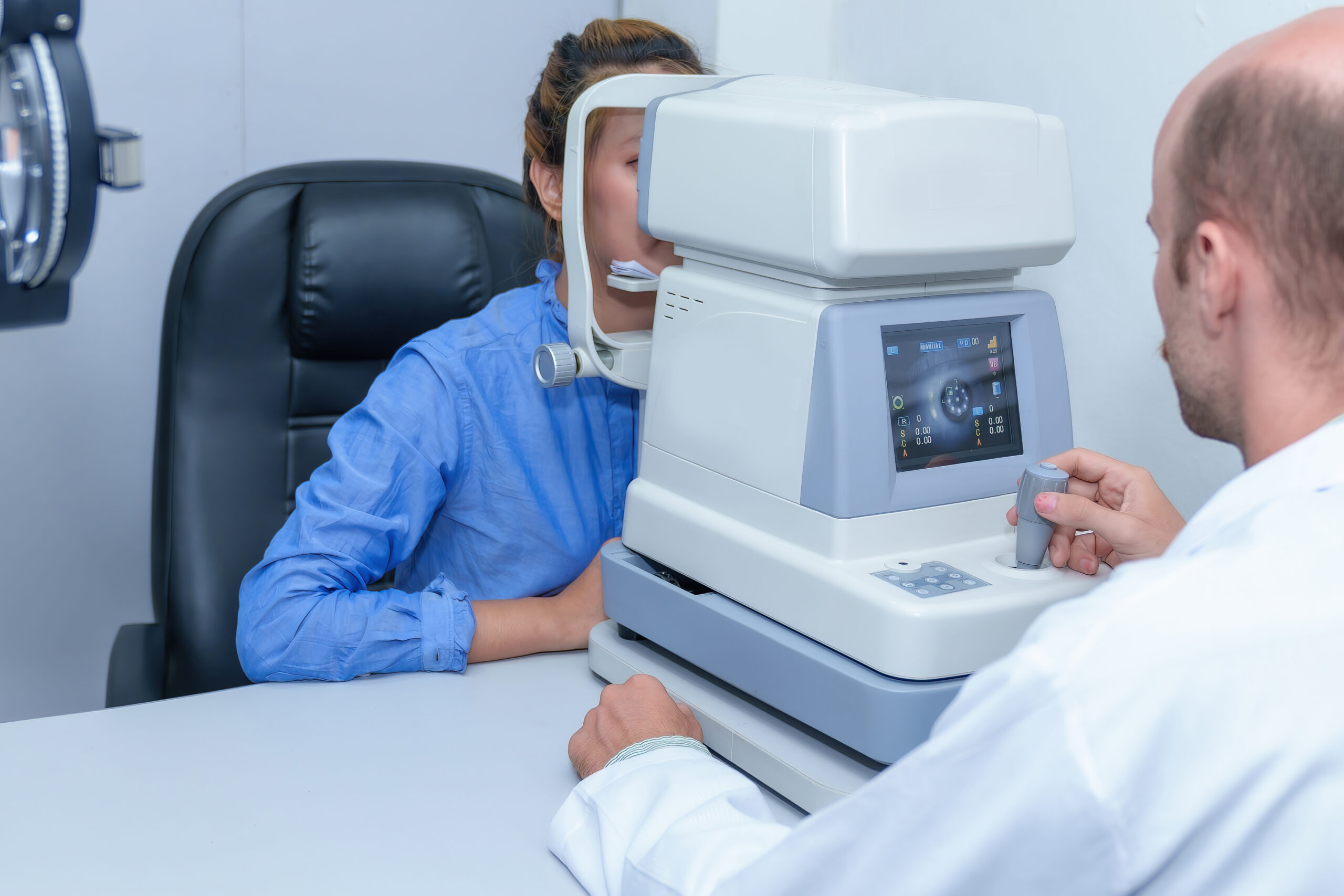
In addition to the basic tests of visual acuity measurement and fundoscopy, many clinicians nowadays consider optical coherence tomography (OCT) imaging to be key for diagnosing and assessing AMD.1 This means equipping your practice with a high-definition OCT is a non-negotiable for those who wish to participate in managing AMD patients. An OCT like the Huvitz HOCT-1/1F with Angiography can be very valuable for the evaluation of wet AMD and neovascular membranes.,3
Discussing AMD with Your Patient
AMD is a relatively well-known condition amongst laypeople; many may associate it with inevitable blindness. This means that receiving a diagnosis of AMD, even without noticeable symptoms, can be devastating for some patients. Conversely, one study reported that patients who were unaffected by visual symptoms were unconcerned about their diagnosis.4
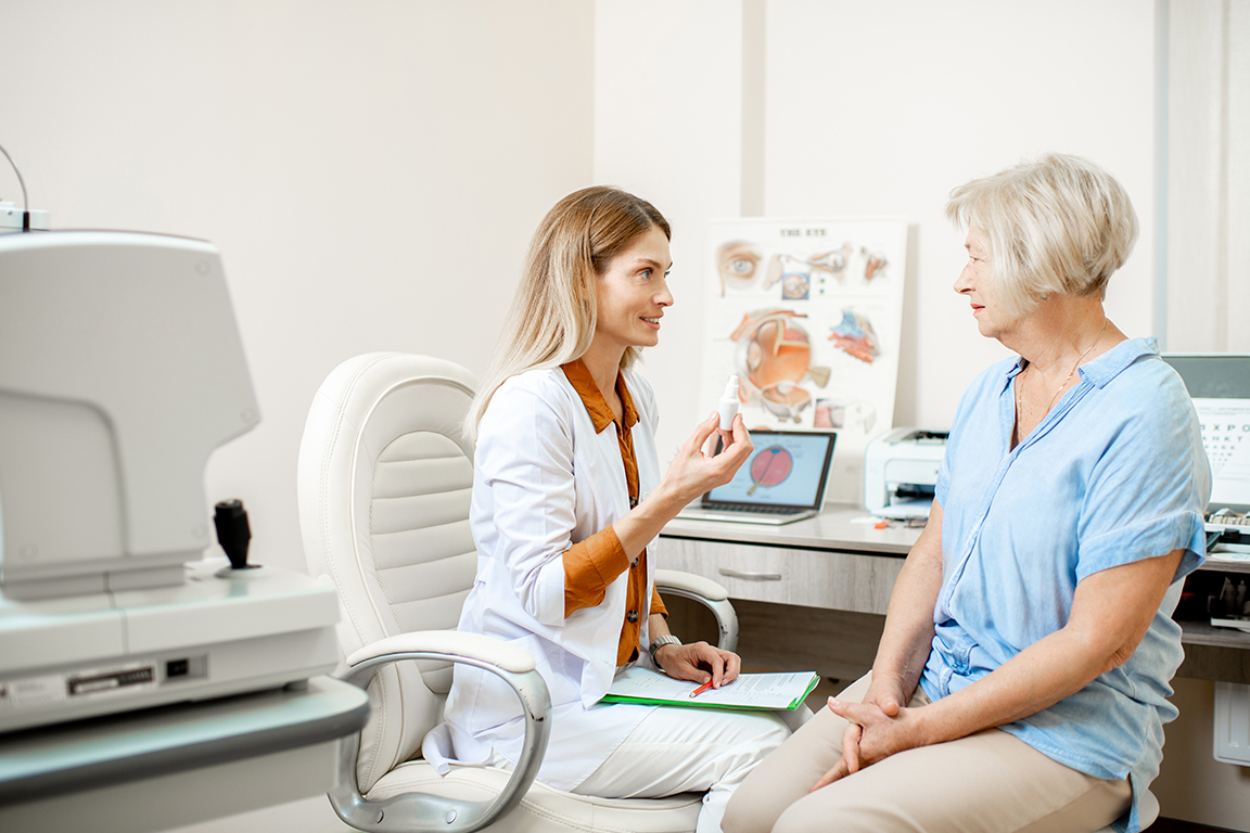
Compassion and impeccable communication are crucial to helping AMD patients navigate their diagnosis and progression of the disease. Successful strategies for breaking the bad news include:4
- Using clear explanations (no medical jargon)
- Outlining the anticipated plan of action for the future
- Taking the time to empathise with the patient
Given that vision impairment from AMD is associated with depression and low mental health,optometrists should also consider being proactive in encouraging their AMD patients to seek counselling or support services. Patients with bilateral geographic atrophy tend to fare worse than those with early or intermediate AMD and those with wet late-stage AMD. This is thought to be due to the availability of treatment or other therapeutic interventions (such as quitting smoking), while historically, there has been no treatment option for late dry AMD.5
Dietary and Lifestyle Advice
Certain nutrients are known to positively impact delaying the progression of dry AMD, and clinicians may find patients (even those without AMD) asking whether they should be taking supplements.
The nutritional supplement supported by the most robust clinical data would be from the ARED trials.1 The outcome of these studies has given us the current AREDS2 formulation, comprised of vitamins C and E, zinc, copper, lutein, and zeaxanthin.1,3 When recommending nutritional supplements for AMD patients, it’s important to note that benefit has been demonstrated only for certain groups, namely those with intermediate or late-stage AMD.1,6 Therefore, patients with no AMD or early stage only should be counselled against using this supplementation. Patients with a fear of developing AMD (such as those with a family history) should also have it explained that these supplements do not prevent AMD from developing in the first place.6
The cessation of smoking has been found to have an even larger beneficial effect on AMD development and progression when compared to dietary supplements, including for those who have not yet developed lesions.1 However, one survey found that optometrists are often hesitant to discuss smoking with their patients despite knowing the impact of this lifestyle choice.7 As primary eyecare providers, it is the optometrist’s role to educate the patient to give them the best opportunity to reduce their risk of disease.
Low Vision Services
Low vision is often considered a more specialised area of optometry, and many optometrists need to be better equipped to help their low vision patients navigate their new experiences. However, this should not prevent clinicians from referring appropriate patients to the low vision services readily available in Australia, such as:
For patients who may not be ready for referral to an external low vision service, you can still offer some simple recommendations for making life a bit easier. This includes tips such as increasing the lighting around the house or for certain tasks, making the most of contrast by pouring light-coloured drinks into dark-coloured cups and vice versa or using smartphones and handheld magnifiers to enlarge print or images.
Summary
As primary eye care providers, optometrists are often the first to become involved in caring for a patient with age-related macular degeneration. It can take extra time and effort to comprehensively assess and manage a person with AMD instead of simply referring them away. However, management of most of these individuals will be well within the scope of an optometrist looking to do the best for their AMD patients.
References
- Stahl A. The Diagnosis and Treatment of Age-Related Macular Degeneration. Dtsch Arztebl Int. 2020; 117(29-30):513-520.
- Optometry Australia. Clinical classification for Age-Related Macular Degeneration (AMD). Pharma. 2019. Available at: https://www.optometry.org.au/wp-content/uploads/Publications/Pharma/2019/Clinical-classification-for-AMD.pdf.
- American Academy of Ophthalmology. Age-Related Macular Degeneration. https://eyewiki.aao.org/. 2022. Available at: https://eyewiki.aao.org/Age-Related_Macular_Degeneration#:~:text=choroidal%20neovascular%20membranes-,Classification,are%20more%20specific%20for%20ARMD). (Accessed May 2023).
- Taylor DJ, Jones L, Binns AM, Crabb DP. ‘You’ve got dry macular degeneration, end of story’: a qualitative study into the experience of living with non-neovascular age-related macular degeneration. Eye (Lond). 2020;34(3):461-473.
- Review of Optometry. Mental Health Issues Linked to AMD, VA. https://www.reviewofoptometry.com/. 2022. Available at: https://www.reviewofoptometry.com/article/mental-health-issues-linked-to-amd-va. (Accessed May 2023).
- NIH National Eye Institute. AREDS/AREDS2 Frequently Asked Questions. https://www.nei.nih.gov/. 2020. Available at: https://www.nei.nih.gov/research/clinical-trials/age-related-eye-disease-studies-aredsareds2/aredsareds2-frequently-asked-questions. (Accessed May 2023).
- Healio. Survey: Optometrists uncomfortable discussing tobacco behaviours with patients. https://www.healio.com/. 2014. Available at: https://www.healio.com/news/optometry/20140715/survey-optometrists-uncomfortable-discussing-tobacco-behaviors-with-patients. (Accessed May 2023).