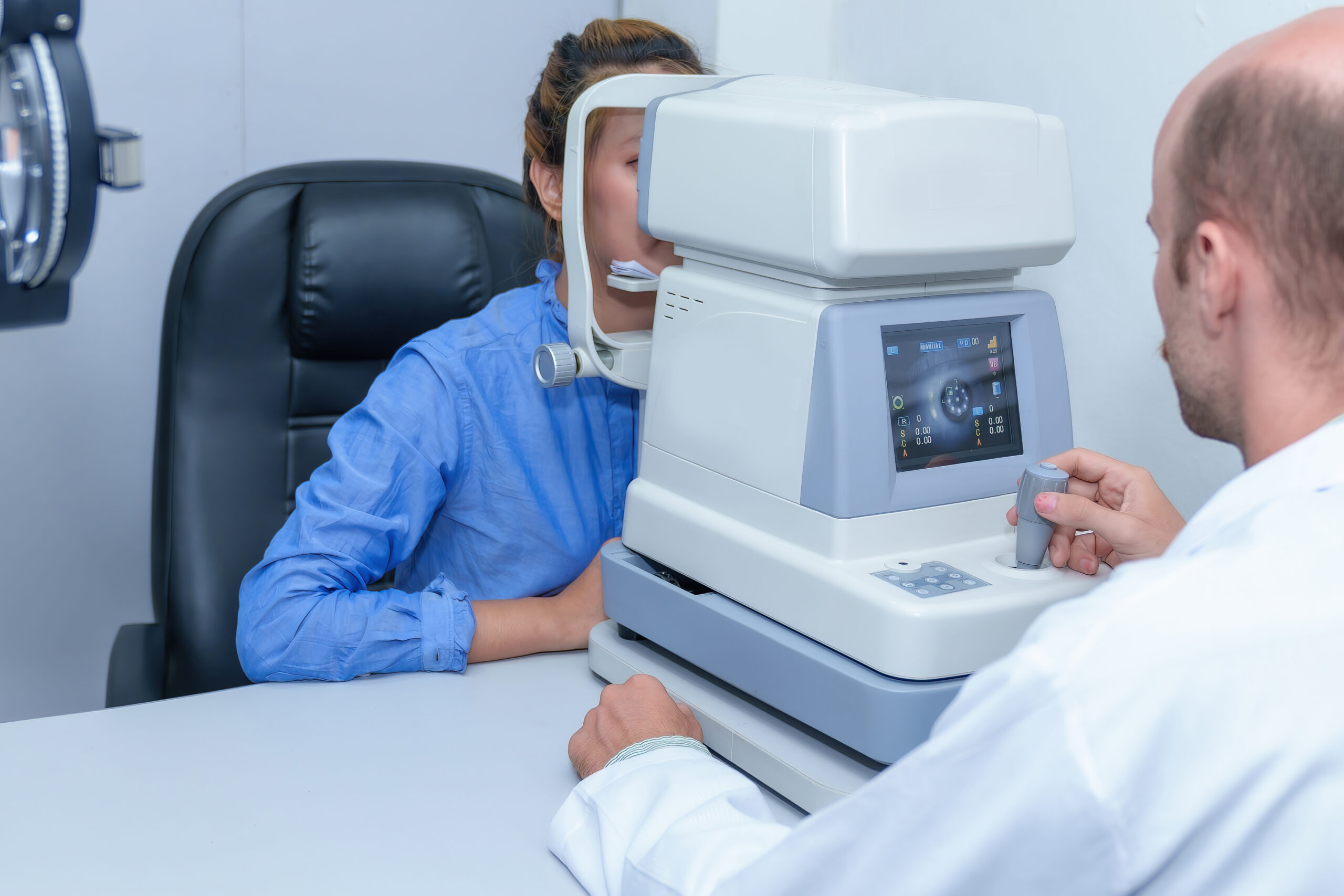
Diagnosing and managing glaucoma is a part of everyday optical practice. Read this article for tips on how to confidently manage your glaucoma patients.
Diagnosing and managing glaucoma is not new to an optometrist’s scope of practice. However, the more we learn about this complex condition, the more complex it seems. Different optometrists will have varying levels of confidence in managing this disease – some will refer to an ophthalmologist in the presence of intraocular pressures (IOP) over 21mmHg, while others are happy to initiate topical therapy and provide long-term follow-up. A comprehensive guide on diagnosing and managing glaucoma is beyond the scope of this article – the NHMRC guidelines are 181 pages! However, here are the essential considerations for confidently managing your glaucoma patients.
Keep yourself up to date.
Our understanding of glaucoma is constantly advancing. For example, neuroprotection as a therapeutic strategy has been of interest for a while, but its success in clinical practice was limited.1 However, more recently, we are seeing promising results from a trial of vitamin B3 (nicotinamide) in slowing the progression of primary open-angle glaucoma.2
Keeping up to date with these developments can help you, as the optometrist, to offer relevant and accurate information to your patients. Helpful resources include those provided by Optometry Australia for members, such as Pharma magazine, the Clinical and Experimental Optometry journal, or articles from Optometry Connection. If you’re feeling so inclined, you may even consider undertaking further education with a Master of Clinical Optometry available at certain universities around Australia. Many ophthalmology clinics will also provide CPD events for optometrists that cover new developments in glaucoma management.
Know when to refer and when to monitor.
Part of this is understanding the limits of your knowledge and abilities. A straightforward example is acute closed-angle glaucoma. Although an optometrist can provide emergency treatment for a patient experiencing an angle closure attack if appropriately equipped, once the patient is stabilised, they must be referred to an ophthalmologist immediately for further treatment.3
The management of open-angle glaucoma may fall into more of a grey area as to whether the patient may be co-managed by an optometrist or referred to the ophthalmologist for exclusive management. This should be decided in collaboration between the patient and the optometrist, even if the optometrist feels confident in their ability. Some patients simply feel more comfortable under a specialist. Remember, optometrists in Australia may independently prescribe topical anti-hypertensives for the treatment of glaucoma provided the patient is referred to an ophthalmologist within four months of initiating treatment.4
The RANZCO recommendations for glaucoma patients who should be exclusively managed by an ophthalmologist include those:5
- With complex ocular pathology
- With secondary glaucoma
- With a history of ocular trauma
- Who are monocular
- Who have undergone multiple ocular procedures
- With advanced glaucoma, including unstable cases or those with IOP >35mmHg
Ensure your practice is equipped appropriately to manage glaucoma.
This is also in line with knowing when to refer and when it’s appropriate to monitor. If your clinic doesn’t have a working visual fields machine, you won’t be able to provide care for your glaucoma patients to an acceptable standard, at least in the long term.
According to the Optometry Australia’s Glaucoma Clinical Practice Guide, there is a minimum requirement in regard to practice equipment for competently managing glaucoma patients.6
- Tonometer, with Goldmann applanation tonometry being the gold standard. Non-contact tonometry (NCT) may also be used though some comparative studies against Goldmann applanation tonometers show they are only reliable if the patient is normotensive.7 Though NCT has other advantages, it may be of limited use in a practice that routinely manages glaucoma patients.
- Pachymeter, either with a handheld pachymeter or an optical coherence tomographer with anterior segment capabilities. Measuring central corneal thickness provides information about this risk factor for primary open angle glaucoma.
- Slit lamp. Needless to say, no optometry practice is complete without a slit lamp. In the context of glaucoma management, this allows viewing of the optic nerve head, retinal nerve fibre layer, and assessment of the anterior chamber angle with gonioscopy.
- Automated threshold visual field machine. There are several different testing algorithms available, for example, SITA Standard or the newer SITA Faster, which have their own pros and cons. A variety of visual field machines are also available, including the VisuScience Hiden LED Arrray Perimeter. Detection and monitoring of visual field loss are crucial for glaucoma management.
Remember, those eyes with glaucoma are attached to a real human.
As eyecare practitioners who see hundreds of glaucoma patients every year, it can be easy to forget the impact this diagnosis can have on a patient. Studies have shown that glaucoma patients have a higher likelihood of mental illness compared to controls, including a higher risk of anxiety and depression.8 There is also evidence that glaucoma can negatively impact a patient’s quality of life.9
Being diagnosed with glaucoma can sometimes be misinterpreted as a diagnosis of complete, inevitable blindness. When first discussing glaucoma with a patient, it’s worthwhile explaining the visual effects of the condition without downplaying the likely prognosis.
If time doesn’t permit a prolonged, in-depth discussion to address all your patient’s concerns (or even if it does), consider directing them to other patient-friendly resources. Understanding more about their diagnosis can help them to feel empowered and reduce anxiety. Glaucoma Australia can be a good resource for patients recently diagnosed with glaucoma.
Are retinal imaging and OCT crucial for glaucoma management?
The current recommendations do not stipulate fundus photography or posterior OCT as part of glaucoma management. However, there is no question that they are extremely valuable in diagnosing early, or pre-perimetric, glaucoma, as well as monitoring disease progression. Depending on your device, OCT scanning may be used to assess peripapillary retinal nerve fibre layer thickness as well as perform a macular ganglion cell count, both of which are useful for glaucoma management.10 If you do intend to depend on OCT scans for glaucoma management, ensure that you’re able to accurately interpret the results to avoid “red and green disease”. Again, investing in furthering your education to get the most out of your OCT benefits you and your patients.
Summary
For most optometrists, diagnosing and managing glaucoma is a part of everyday practice. Ensuring both you as an eyecare practitioner and your practice is well-equipped to provide excellent care for your glaucoma patients can take some thought and investment of time, money, and effort. However, the satisfaction from providing high-quality care for your patients is more than worthwhile.
References
- Glaucoma Today. Neuroprotection and Glaucoma: Old Challenges and New Solutions. Available at: https://glaucomatoday.com/articles/2019-nov-dec/neuroprotection-and-glaucoma-old-challenges-and-new-solutions. (Accessed December 2022).
- CERA. Follow-up trial to determine role of vitamin B3 in glaucoma treatment. Available at: https://www.cera.org.au/vitamin-b3-trial-to-determine-its-role-in-glaucoma-treatment/#:~:text=%E2%80%9CAs%20a%20safe%20therapy%20that,been%20damaged%20to%20function%20better. (Accessed December 2022).
- Murray D. Emergency management: angle-closure glaucoma. Community Eye Health. 2018;31(103):64.
- Optometry Australia. Sharing the care for glaucoma. 2016. Available at: https://www.optometry.org.au/workplace/sharing-the-care-for-glaucoma/. (Accessed December 2022).
- RANZCO. Clinical Practice Guidelines for the Collaborative Care of glaucoma patients and suspects by ophthalmologists and optometrists in Australia. 2019. Available at: https://ranzco.edu/wp-content/uploads/2018/11/Guidelines-for-the-Collaborative-Care-of-Glaucoma-Patients.pdf.
- Optometry Australia. Clinical Practice Guide for the Diagnosis and Management of Open Angle Glaucoma. 2020. Available at: https://www.optometry.org.au/wp-content/uploads/Professional_support/Guidelines/Glaucoma-Clinical-Practice-Guide_Dec-2020_design_v5.pdf.
- Shah M, Saleem K, Mehmood T. Intraocular Pressure Measurement: Goldmann Applanation Tonometer vs Non Contact Air Puff Tonometer. J Ayub Med Coll Abbottabad.2012;24:3-4.
- Ubochi CC, Achigbu EO, Nkwogu FU, Onyia OE, Okeke CJ. The Impact of Glaucoma on the Mental Health of Primary Open-Angle Glaucoma Patients Attending a Teaching Hospital in South East Nigeria. J West Afr Coll Surg. 2020;10(2):17-22.
- Zhang, Q., Zhou, W., Song, D. et al. Vision-related quality of life in patients with glaucoma: the role of illness perceptions. Health Qual Life Outcomes.2022;20:78.
- Review of Ophthalmology. Monitoring Glaucoma Progression with OCT. 2020. Available at: https://www.reviewofophthalmology.com/article/monitoring-glaucoma-progression-with-oct. (Accessed December 2022).


